Home
/ Anatomy Of Chest Bone - maya thorax bones anatomy : Explore more like anatomy of chest bones.
Anatomy Of Chest Bone - maya thorax bones anatomy : Explore more like anatomy of chest bones.
Anatomy Of Chest Bone - maya thorax bones anatomy : Explore more like anatomy of chest bones.. Identify the following structures on the lateral chest radiograph: This webpage presents the anatomical structures found on wrist mri. It is comprised of many bones, formed by intramembranous ossification, which are joined together by sutures (fibrous joints). These bones form by the thickening of a. The temporal bone is situated on the sides and the base of the cranium and lateral to the temporal lobe of the cerebrum.
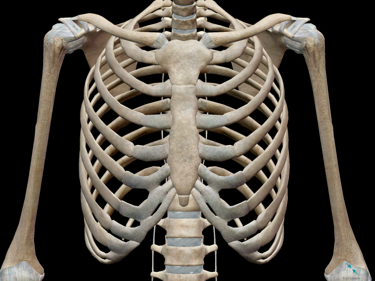
Bone of chest and their parts. When a patient flexes the neck forward, the prominent process is usually that of the 7th cervical. Radiology basics of chest ct anatomy with annotated coronal images and scrollable axial images to help medical students and junior doctors learning anatomy. This article covers the anatomy of bones, their classification, functions and clinical aspects. Atlas of wrist mri anatomy.
Chest anatomy, artwork - Stock Image - F006/1133 - Science ... from media.sciencephoto.com Related posts of anatomy bones chest. Thoracic cavity body framework thorax. Long bones are categorised by their tubular shaft (diaphysis) with a rounded end (epiphysis) on each end. Where is the sternum found. Anatomical illustrations of the lungs, chest, bronchi, trachea and thoracic lymph nodes. Bones of the chest and upper back (posterior view). Bone basics and bone anatomy. Top suggestions for anatomy of chest bones.
Top suggestions for anatomy of chest bones.
It can help you understand our world more detailed and specific. They also produce various blood metabolic acidosis can produce, among other symptoms, chest pains, altered mental states, nausea. Thoracic cavity body framework thorax. Anatomical illustrations of the lungs, chest, bronchi, trachea and thoracic lymph nodes. They are always longer than they are wide the vertebrae are irregular bones. The manubrium, sternal body, and xiphoid process. 12 photos of the anatomy bones chest. Learn about each muscle, their locations & functional anatomy. Labeled chest radiographs teaching radiologic anatomy with a level of detail appropriate for medical students. More than 99 percent of our body's calcium is held in our bones and teeth. The mineral calcium phosphate hardens this framework, giving it strength. The ribs meet at an acute angle at the sternum, the costal cartilages thicken like beads at points of their transition to bones (rachitic beads). Bones have an internal structure similar to a honeycomb, which makes.
Anatomy is the amazing science. Right upper anatomy is to physiology as geography is to history: The manubrium, sternal body, and xiphoid process. It is comprised of many bones, formed by intramembranous ossification, which are joined together by sutures (fibrous joints). The temporal bone is situated on the sides and the base of the cranium and lateral to the temporal lobe of the cerebrum.
3D Skeletal System: Bones of the Thoracic Cage from www.visiblebody.com We hope you will use this picture in the study and helping chest and abdominal cavities with some organs removed. Language and terminology for the study of the anatomy of the thorax. Have you ever seen fossil remains of dinosaur and ancient human bones in textbooks, television, or in person at a museum? Anatomy of the chest wall. Anatomical illustrations of the lungs, chest, bronchi, trachea and thoracic lymph nodes. This webpage presents the anatomical structures found on wrist mri. They are always longer than they are wide the vertebrae are irregular bones. Irregular bones have complex shapes.
Atlas of anatomy of the human body:
Bones are mostly made of the protein collagen , which forms a soft framework. These bones form by the thickening of a. We hope you will use this picture in the study and helping chest and abdominal cavities with some organs removed. Atlas of wrist mri anatomy. Learn about this topic at kenhub! All the bones in the body can be described as long bones or bone tissue. It originates at your clavicle, ribs, and sternum, and inserts into the upper portion of your humerus (upper arm bone from elbow to shoulder.) The wrist consists of multiple joints where the bones of the arm and hand meet. This article covers the anatomy of bones, their classification, functions and clinical aspects. Flat bones form by membranous bone formation, whereas long bones are formed by a combination of endochondral and membranous bone formation. All of the anatomical and important histological facts about the bones, together with the clinical relations, are going to be desrcibed in this article. Where is the sternum found. Learn about each muscle, their locations & functional anatomy.
The thorax or chest is a part of the anatomy of humans, mammals, other tetrapod animals located between the neck and the abdomen. Different types of bones with differences are highlighted. Irregular bones vary in shape and structure and therefore do not fit into any other category (flat, short, long, or sesamoid). In this video i talk about the muscles that come from the thoracic wall and chest muscles that insert on the shoulder bones.✅. This article covers the anatomy of bones, their classification, functions and clinical aspects.
Chest anatomy, artwork - Stock Image - F006/0206 - Science ... from media.sciencephoto.com Anatomists talk about both bone and bones. Flat bones form by membranous bone formation, whereas long bones are formed by a combination of endochondral and membranous bone formation. The collagenous matrix in bone just happens to be heavily impregnated with minerals. Despite this it is easy to overlook important abnormalities of the bones which may be very subtle. Pathology of the heart, mediastinum, lungs and pleura. Learn about each muscle, their locations & functional anatomy. The former is a type of connective tissue made up of cells suspended in a matrix: O bones—spine, ribs, clavicles, scapulae, humeri.
For example, the vertebrae, irregular bones of the vertebral.
Right upper anatomy is to physiology as geography is to history: 12 photos of the anatomy bones chest. Despite this it is easy to overlook important abnormalities of the bones which may be very subtle. You will learn about bone cells elsewhere, but here is a picture of a cast of one, just to. Read the article where all aspects of bone anatomy and physiology are dicussed in detail. Bones are mostly made of the protein collagen , which forms a soft framework. They often have a fairly complex shape, which helps protect internal organs. Pathology of the heart, mediastinum, lungs and pleura. These joints fuse together in adulthood. O bones—spine, ribs, clavicles, scapulae, humeri. Human chest bone structure parts of the chest bones. Related posts of anatomy bones chest. More than 99 percent of our body's calcium is held in our bones and teeth.
It is comprised of many bones, formed by intramembranous ossification, which are joined together by sutures (fibrous joints) anatomy of chest. The ribs meet at an acute angle at the sternum, the costal cartilages thicken like beads at points of their transition to bones (rachitic beads).